
Aignostics Brings Tertiary Lymphoid Structures and Whole Tumor Regions to Atlas H&E-TME and OpenTME
New spatial features add organized immune architecture and tumor context to AI-powered spatial profiling

We're excited to announce two new spatial features for Atlas H&E-TME, our AI-powered application for tumor microenvironment (TME) analysis: tertiary lymphoid structure (TLS) detection and whole tumor region (WTR) detection. TLSs are immune structures whose presence and maturity track with immunotherapy response in cancer; WTR identifies the tumor itself, from its core to the invasive margin, the frame of reference for the wider microenvironment. Both are also available in OpenTME, our open-access dataset of thousands of TCGA whole-slide images.
Assessing TLSs to predict response to therapy
Predicting who will respond to checkpoint-inhibitor therapy remains one of immuno-oncology's central challenges, and TLSs have emerged as a promising spatial signal: their presence, and especially their maturity, tracks with response and better prognosis across a range of solid tumors. Much of this is visible on H&E to a trained eye, but assessing every structure across a cohort by hand is slow manual work.
Atlas H&E-TME can assess TLSs from routine H&E slides alone, identifying each one, placing it in its tissue context, and returning analysis-ready readouts at the structure and slide level. It also classifies each by maturity, from immature aggregates to fully germinal-center–bearing forms, and summarizes them by class. Few automated tools reach that resolution, and maturity is the property most closely tied to clinical outcomes.

Mapping WTR to read the microenvironment in context
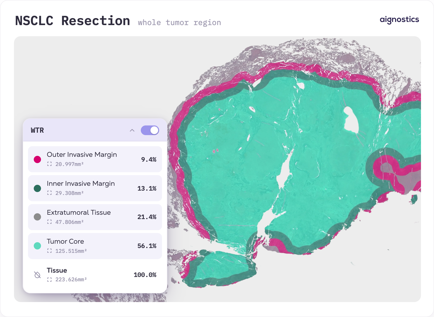
Location determines meaning: a signal inside the tumor is not the same as one in distant benign tissue. WTR detection maps the tumor and provides a reference for the immune features measured within and around it. Our new WTR feature for Atlas H&E-TME resolves each slide from the tumor core out to the surrounding extratumoral tissue, distinguishing the inner and outer invasive margin in between, so readouts can be reported for each. These regions have traditionally been drawn by expert annotation; automation brings the same delineation to entire cohorts, more quickly and consistently.
With the tumor delineated from surrounding tissue, features like immune phenotype (inflamed / desert / excluded), tumor-infiltrating lymphocytes, and vascularization can be assessed within the tumor itself rather than across the whole slide.
Getting started
Both features come as standard outputs: polygon overlays viewable in the Aignostics pathology viewer or your own image management system, plus exportable quantitative readouts. They join our industry-leading capabilities, such as quality control, tissue segmentation, cell classification, and quantitative readouts. In a recent preprint, Atlas H&E-TME matched or exceeded expert pathologists against an IHC-informed consensus. TLS and WTR are available across every indication Atlas H&E-TME supports.
For life science teams, the features will help address key immuno-oncology questions, such as how TLS maturity and localization relate to treatment response or survival, or how the tumor's immune landscape shifts under treatment. These analyses can also be extended to archival cohorts where specialized staining was never performed. Teams can request a free trial on our website.
Academic researchers can explore both features in OpenTME, where they come pre-computed across thousands of TCGA slides, ready to correlate with molecular and clinical data. Those with their own cohorts can apply to the Research Access Program to run H&E slides through Atlas H&E-TME at no cost.
TLS and WTR are the latest additions to Atlas H&E-TME and OpenTME, which we're continually expanding with new spatial features and broader indication coverage.